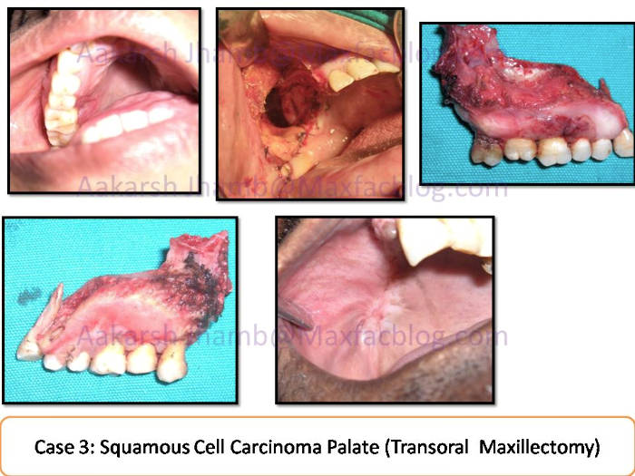
It was one of my earlier patients as Consultant Maxillofacial Surgeon. This 40year old man was referred by a Dentist friend for the chief complaint of Non Healing Ulcer on palate. The patient gave history of Tabacco Chewing and Smoking since 7 years. Clinically it was a superficial ulcer on the Palatal Gingiva of right maxillary molar. Incisional Biopsy showed Well Differentiated Squamous Cell Carcinoma. Contrast CT scan (skullbase to Clavicle) was advised for staging and Preop stage of T2N0M0 (clinical & CT) was done.
Transoral Inferior Maxillectomy was done under General Anesthesia. Neck dissection was not advised because of N0 status and low risk of occult metastasis. No reconstruction was done and only Interim Obturator appliance was inserted in the Maxillary defect. Final Histopathology showed clear margins all more than 1cm from ulcer edge T2N0M0 (Pathologic staging). No Radiation or Chemotherapy was advised. The defect closed off completely by 4 months post surgery visit. Partial Removable Denture was made to complete the dental rehabilitation.
This is one of the rare instances when the patient could be diagnosed and treated early, mainly because their dentist was vigilant and wise enough to send them for biopsy. It highlights the vital role which dental surgeons can play in catching early oral malignant lesions.